Категория: Новости.
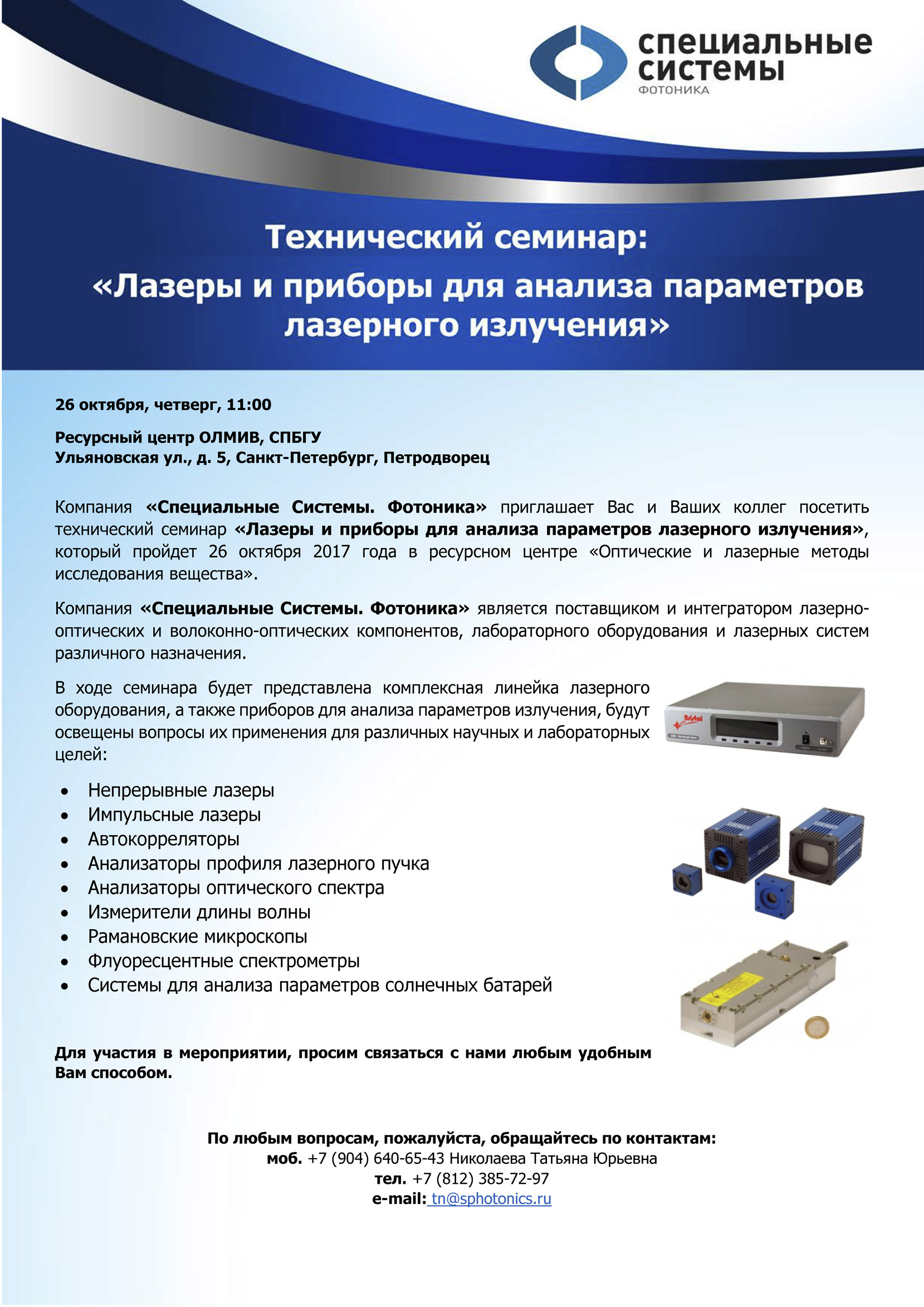
26 октября, четверг, 11:00 в РЦ ОЛМИВ состоится семинар от Компании «Специальные Системы. Фотоника» на тему: «Лазеры и приборы для анализа параметров лазерного излучения». Приглашаются все желающие.

Категория: Новости.
Journal of Molecular Structure Volume 1146, 15 October 2017, Pages 554–561
Anastasiia M. Afanasenko, Dina V. Boyarskaya, Irina A. Boyarskaya, Tatiana G. Chulkova, Yakov M. Grigoriev, Ilya E. Kolesnikov, Margarita S. Avdontceva, Taras L. Panikorovskii, Andrej I. Panin, Anatoly N. Vereshchagin, Michail N. Elinson
Structures and photophysical properties of 3,4-diaryl-1H-pyrrol-2,5- diimines and 2,3-diarylmaleimides
Journal of Molecular Structure Volume 1146, 15 October 2017, Pages 554–561
DOI: 10.1016/j.molstruc.2017.06.048

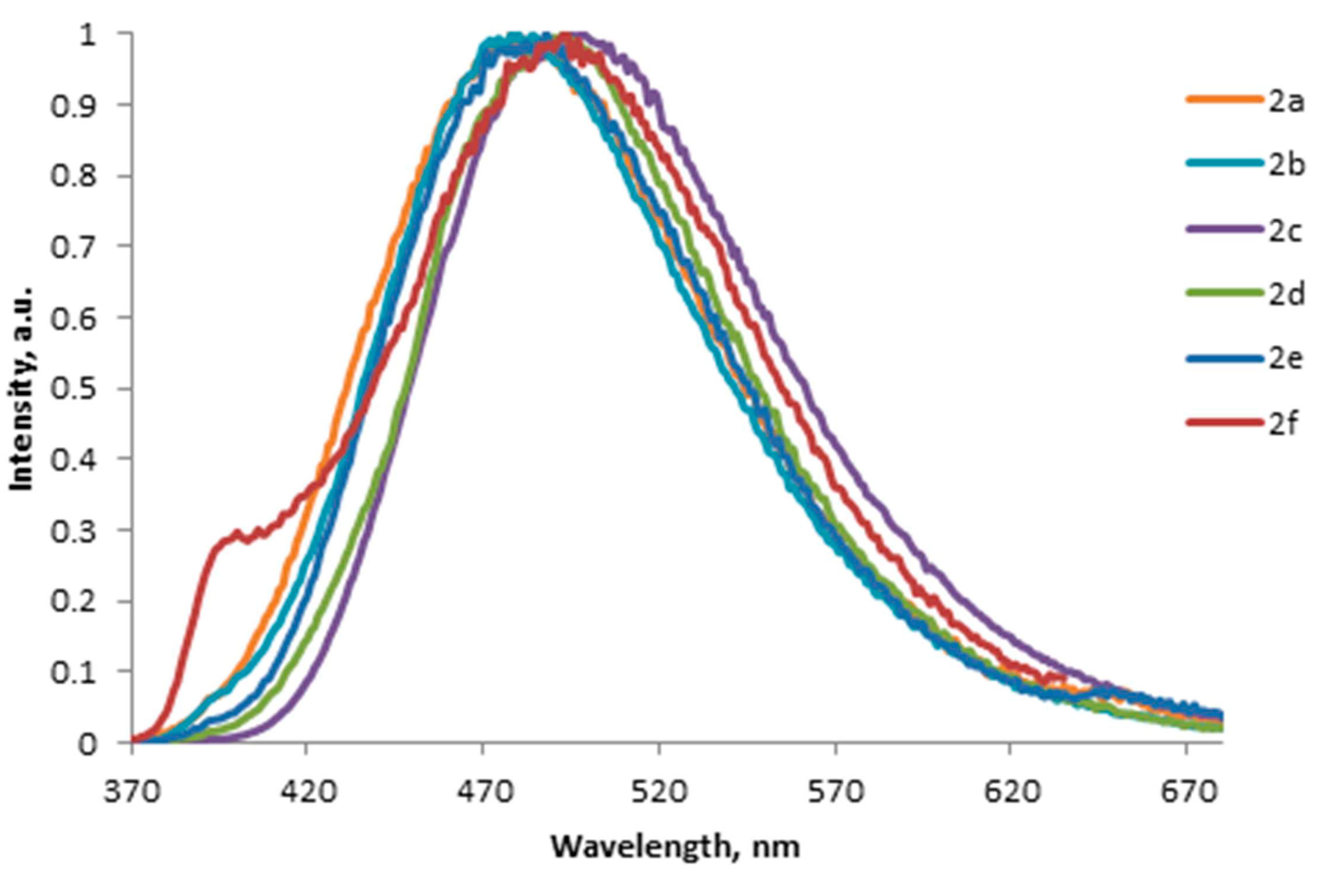
Structural features of 3,4-diaryl-1H-pyrrol-2,5-diimines and their derivatives have been studied by molecular spectroscopy techniques, single-crystal X-ray diffraction, and DFT calculations. According to the theoretical calculations, the diimino tautomeric form of 3,4-diaryl-1H-pyrrol-2,5-diimines is more stable in solution than the imino-enamino form. We also found that the structurally related 2,3-diarylmaleimides exist in the solid state in the dimeric diketo form. 3,4-Diaryl-1H-pyrrol-2,5-diimines and 2,3-diarylmaleimides exhibit fluorescence in the blue region of the visible spectrum. The fluorescence spectra have large Stokes shifts. Aryl substituents at the 3,4-positions of 1H-pyrrol-2,5-diimine do not significantly affect fluorescence properties. The insertion of donor substituents into 2,3-diarylmaleimides leads to bathochromic shift of emission bands with hyperchromic effect.
Категория: Новости.
Tsitologiya Volume 59, Issue 10, Pages 662–668
V. A. Kulichkova, A.V. Selenina, A. N. Tomilin, A.S. Tsimokha
ESTABLISHMENT OF A HELA CELL LINE, STABLY EXPRESSING EXOSOME MARKER CD63 FUSED WITH THE FLUORESCENT PROTEIN TagRFP AND HTBH TAG
Tsitologiya Volume 59, Issue 10, Pages 662–668
Экзосомы - микроскопические внеклеточные везикулы диаметром 30-100 нм, выделяемые в межклеточное пространство различными клетками млекопитающих. Эти везикулы обнаруживаются в большом количестве в сыворотке крови и других внеклеточных жидкостях организмов и содержат большой набор разнообразных белков, мРНК и микроРНК. Экзосомы участвуют в межклеточной коммуникации, секреции белков, иммунном ответе, а также вовлечены в развитие нейродегенеративных и онкологических заболеваний. Механизмы, с помощью которых они выходят или поступают в клетки, недостаточно изучены. В настоящей работе создана стабильная клеточная линия на основе клеток аденокарциномы шейки матки человека HeLa, экспрессирующая маркер экзосом, тетраспанин CD63, слитый с красным флуоресцентным белком TagRFP и последовательностью HTBH. Полученная линия клеток HeLa, экспрессирующая белок CD63-TagRFP-HTBH, позволит наблюдать за транспортом экзосом прижизненно.
Категория: Новости.
Applied Surface Science Volume 416, September 2017, Pages 988-995
A.A. Ionina, S.I. Kudryashova, A.O. Levchenko, L.V. Nguyen, I.N. Saraev, A.A. Rudenko, E.I. Ageev, D.V. Potorochin, V.P. Veiko, E.V. Borisov, D.V. Pankin, D.A. Kirilenko, P.N. Brunkov
Correlated topographic and structural modification on Si surface during multi-shot femtosecond laser exposures: Si nanopolymorphs as potential local structural nanomarkers
Applied Surface Science Volume 416, September 2017, Pages 988-995
DOI: 10.1016/j.apsusc.2017.04.215

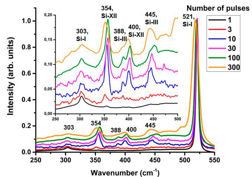
High-pressure Si-XII and Si-III nanocrystalline polymorphs, as well as amorphous Si phase, appear consequently during multi-shot femtosecond-laser exposure of crystalline Si wafer surface above its spallation threshold along with permanently developing quasi-regular surface texture (ripples, microcones), residual hydrostatic stresses and subsurface damage, which are characterized by scanning and transmission electron microscopy, as well as by Raman micro-spectroscopy. The consequent yields of these structural Si phases indicate not only their spatially different appearance, but also potentially enable to track nanoscale, transient laser-induced high-pressure, high-temperature physical processes - local variation of ablation mechanism and rate, pressurization/pressure release, melting/resolidification, amorphization, annealing - versus cumulative laser exposure and the related development of the surface topography.
Категория: Новости.
Optics and Lasers in Engineering Volume 96, September 2017, Pages 63-67
Vadim Veiko, Yulia Karlagina, Mikhail Moskvin, Vladimir Mikhailovskii, Galina Odintsova, Pavel Olshin, Dmitry Pankin, Valery Romanov, Roman Yatsuk
Metal surface coloration by oxide periodic structures formed with nanosecond laser pulses
Optics and Lasers in Engineering Volume 96, September 2017, Pages 63-67
DOI: 10.1016/j.optlaseng.2017.04.014

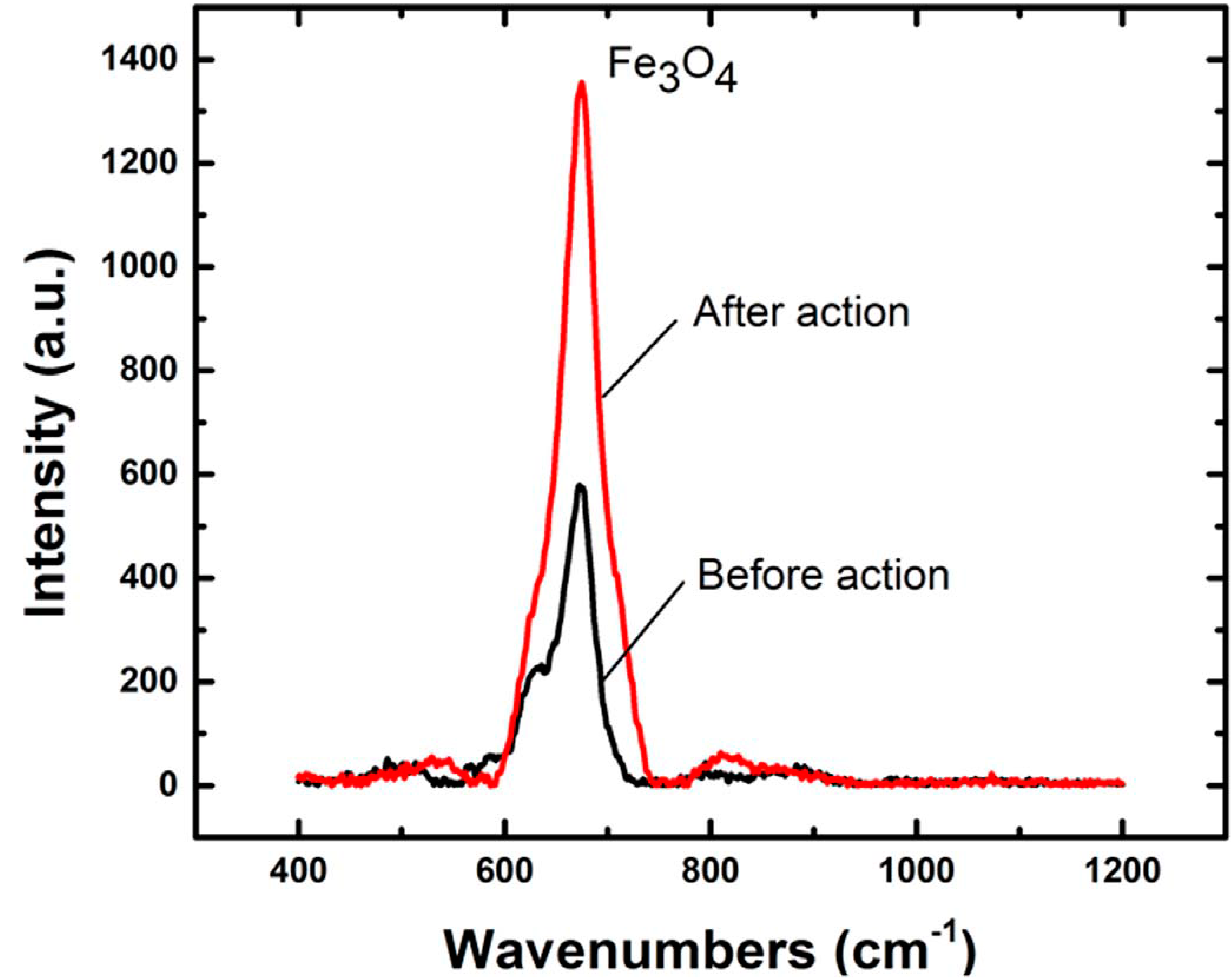
In this work, we studied a method of laser-induced coloration of metals, where small-scale spatially periodic structures play a key role in the process of color formation. The formation of such structures on a surface of AISI 304 stainless steel was demonstrated for the 1.06 μm fiber laser with nanosecond duration of pulses and random (elliptical) polarization. The color of the surface depends on the period, height and orientation of periodic surface structures. Adjustment of the polarization of the laser radiation or change of laser incidence angle can be used to control the orientation of the structures. The formation of markings that change their color under the different viewing angles becomes possible. The potential application of the method is metal product protection against falsification.


 Русский (РФ)
  English (UK)

