Категория: О ресурсном центре.
Научно-техническое исследование объектов культурного наследия
Более 10 лет на базе РЦ ОЛМИВ (ранее в НИИ лазерных исследований) ведется работа в направлении научно-технического исследования объектов культурного наследия. Квалификация сотрудников центра, имеющих преимущественно высшее физическое химическое образование позволяет детально прорабатывать задачи по изучению материальной основы памятников, а плотное взаимодействие с реставраторами, искусствоведами и хранителями ведет к успешному выполнению междисциплинарных работ.
Ресурсный центр проводит анализ вещества с использование следующих оптических методов:
- ИК-Фурье спектроскопия поглощения,
- спектроскопия поглощения УФ/видимого/ближнего ИК диапазона,
- спектроскопия комбинационного рассеяния света (микро-Рамановская спектроскопия),
- спектроскопия люминесценции,
- исследовательская фотография.
- лазерная чистка.
Кроме того, в центре имеется возможность предварительной пробоподготовки образцов.
В разные годы РЦ ОЛМИВ проводил исследования совместно с такими организациями, как
- Факультет искусств СПбГУ
- Государственный Эрмитаж
- Санкт-Петербургский государственный академический институт живописи, скульптуры и архитектуры имени И.Е. Репина при Российской академии художеств
- Библиотека Российской Академии Наук
- Лаборатория консервации и реставрации Архива РАН
- Санкт-Петербургская государственная художественно-промышленная академия имени А. Л. Штиглица
Mie scattering meets interferometry at the nanoscale – Resolving the vectorial nature of light from the far‐field.
Prof. Gerd Leuchs, Max Planck Institute for the Science of Light, Erlangen, Germany
When light gets tightly focused down to a size of the order of the wavelength, it exhibits a complex electromagnetic field distribution with deep sub‐wavelength features. Such highly confined light fields find a wide range of applications in microscopy, imaging and nanooptics, where achievable resolution and the information content of an investigated system strongly depend on the precise knowledge of the illuminating light fields. However, measuring their full vectorial distribution is not an easy task, especially in the diffraction‐limited focal spot of a high numerical aperture system.
We now developed an experimental nanoprobing scheme, called ‘Mie scattering nanointerferometry’, which allows for the measurement of both amplitude and phase distributions of the electric field in highly confined light with nanometer resolution. We extract this information from the scattering signal of a single nanoparticle with sub‐wavelength dimensions utilized as a field probe. By simply measuring and analyzing the scattering pattern recorded in the far‐field for different positions of the particle relative to the probed field, the distributions of the individual electric field components can be experimentally reconstructed. We take advantage of the interference between the incoming focused light beam and the light scattered off the nanoprobe. The phase information of the investigated field gets stored in this interference term, which is naturally included in the chosen system, and can be recovered by the abovementioned spatially and angularly resolved far‐field measurement. No complex analysis of the scattered light field in terms of its polarization is required.
This high resolution technique is easy‐to‐implement and highly compatible with standard microscopy systems. It paves the way for a highly precise and accurate characterization of optical systems. In addition, we utilize this method to investigate novel states of the light field and beam‐shift phenomena at the nanoscale. Furthermore, we believe that it will help to improve existing imaging and microscopy methods, such as laser scanning microscopy. Moreover, it can be adapted to allow for the measurement of even purely evanescent fields.
S. Quabis et al., “Focusing light to a tighter spot,” Opt. Comm., 179, 1 (2000)
P. Banzer et al., “On the experimental investigation of the electric and magnetic response of a single nano‐ structure,” Opt. Express, 18, 10905 (2010)
T. Bauer, S. Orlov, U. Peschel, P. Banzer and G. Leuchs, „Nanointerferometric amplitude and phase reconstruction of tightly focused vector beams,” Nature Photon., 8, 23–27 (2014)
P. Banzer, M. Neugebauer, A. Aiello, C. Marquardt, N. Lindlein, T. Bauer and G. Leuchs, “The photonic wheel ‐ demonstration of a state of light with purely transverse angular momentum,” J. Europ. Opt. Soc. Rap. Public. 8, 13032 (2013)
M. Neugebauer, P. Banzer, T. Bauer, S. Orlov, N. Lindlein, A. Aiello and G. Leuchs, “Geometric spin Hall effect of light in tightly focused polarization tailored light beams,” to be published in Physical Review A (2014)
Категория: О ресурсном центре.
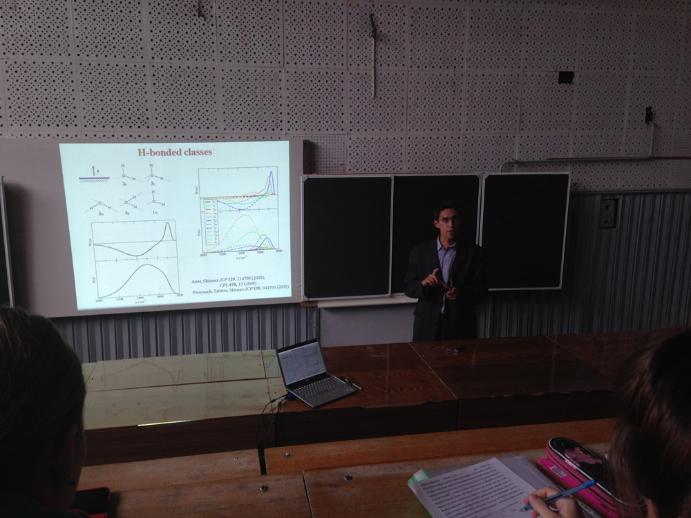
12 ноября 2013 в аудитории Химического факультета 04 состоялся научный семинар «Лазерные методы в химии».
Программа научного семинара:
Доклад А. Бендерского, Департамент химии, Университета Южной Калифорнии, Лос-Анджелес, США
Структура и динамика водородных связей на поверхности воды
Доклад А. Поволоцкого, Кафедра лазерной химии и лазерного материаловедения, Химического факультета СПбГУ
Оборудование и научные исследования в РЦ «Оптические и лазерные методы исследования вещества»
Доклад А. Маньшиной, Кафедра лазерной химии и лазерного материаловедения, Химического факультета СПбГУ
Работы по лазерному материаловедению на кафедре ЛХЛМ
Категория: О ресурсном центре.
11 сентября 2013 года в Ресурсном центре «Оптические и лазерные методы исследования вещества» состоялся семинар INTERTECH Corporation "Современные приборы для ИК и Раман спектроскопии". Компанию INTERTECH представляли старший специалист по оборудованию для ИК-Фурье и КР спектроскопии по Европе, Ближнему Востоку и Африке Бруно Беккард (Bruno J. Beccard) (слева на фото) и технический специалист по оборудованию для ИК и КР спектроскопии Сергей Вадимович Тихомиров (справа на фото).
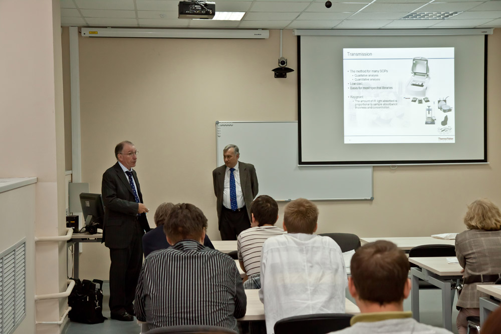
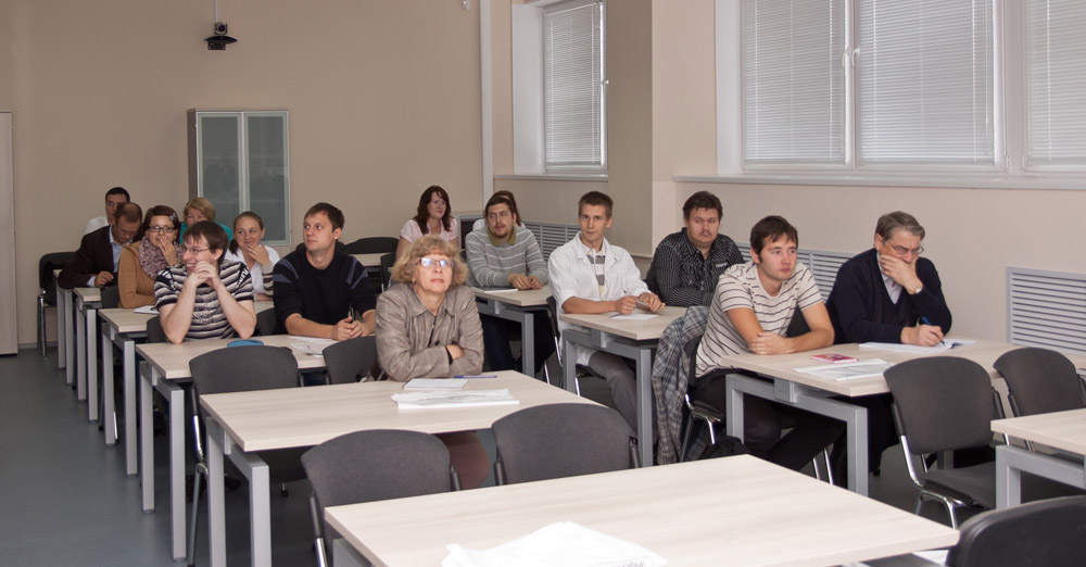
Семинар был посвящен обзору современных ИК-Фурье спектрометров производства Thermo Scientific, инфракрасных микроскопов Thermo Scientific и их применению, а так же были рассмотрены вопросы применения ИК-Фурье спектрометров. В семинаре приняли участие сотрудники Химического, Физического и Биологического факультетов СПбГУ, а так же сотрудники Института цитологии РАН (ИНЦ РАН)

 Русский (РФ)
  English (UK)

